Home
/ Muscles Of The Torso Posterior : Digital Illustration Of Muscles Of The Human Torso Anterior View Stock Photo Alamy : Human muscles · august 21, 2016.
Muscles Of The Torso Posterior : Digital Illustration Of Muscles Of The Human Torso Anterior View Stock Photo Alamy : Human muscles · august 21, 2016.
Muscles Of The Torso Posterior : Digital Illustration Of Muscles Of The Human Torso Anterior View Stock Photo Alamy : Human muscles · august 21, 2016.. Quizzes on the muscles of the torso. L6 drawing the male torso posterior view part 1: The front fibers flex the arm and the middle fibers help abduct the arm (bring the arm away from the body). It is controlled by the axillary nerve. It is attached to the calcaneus and is pulled by 3 flexor muscles:
Pectoralis major, pectoralis minor, serratus anterior and subclavius.collectively, these muscles are involved in movement and stabilisation of the scapula, as well as movements of the upper limb. Muscle anatomy of the human body. We are looking at normans (model) muscles in this video. Adductor magnus biceps femoris carpi flexor ulnaris deltoid. The primary back muscles are divided into a total of five (5) systems, each system comprising medial and lateral tracts of various muscles.
Digital Illustration Of Muscles Of The Human Torso Anterior View Stock Photo Alamy from c8.alamy.com An extremely strong tendon attached to the heel. It inserts onto the inferior border of the 12th rib. Usually as one muscle contracts (or shortens), the opposing muscle (known as the antagonist) elongates and vice versa. Posterior muscles in the body. These muscles are also known as erector spinae (spinal erectors) or erector trunci (truncal erectors) since they specifically describe the primary function: All muscles maintain some amount of muscle tone at all times, unless the muscle has been disconnected from the central nervous system due to nerve damage. In this lesson, we will identify and draw the superficial and deep muscles of the front and rear torso. Pectoralis major, pectoralis minor, serratus anterior and subclavius.collectively, these muscles are involved in movement and stabilisation of the scapula, as well as movements of the upper limb.
L6 drawing the male torso posterior view part 1:
The body has three types of muscles: Skeletal muscle, posterior, unlabeled figure 8.16 250. Chart of major posterior muscles. The muscles of the upper limb can be divided into 6 different regions: Pectoralis major, pectoralis minor, serratus anterior and subclavius.collectively, these muscles are involved in movement and stabilisation of the scapula, as well as movements of the upper limb. This muscle originates from the iliac crest and iliolumbar ligament. Human muscle system, the muscles of the human body that work the skeletal system, that are under voluntary control, and that are concerned with movement, posture, and balance.broadly considered, human muscle—like the muscles of all vertebrates—is often divided into striated muscle (or skeletal muscle), smooth muscle, and cardiac muscle.smooth muscle is under involuntary control and is. There are 4 muscles of the pectoral region: Enjoy a selection of illustrations, sketches, model sheets and tutorials by various artists, collected and shown here for educational and inspirational. To learn about muscles effectively, a clear and logical grouping into systems with unique structures is needed. Collection by character design references. An extremely strong tendon attached to the heel. Chest muscles function in respiration while abdominal muscles function in torso movement and in maintenance of balance and posture muscles of torso.
It is attached to the calcaneus and is pulled by 3 flexor muscles: Posterior muscles of the torso : It is controlled by the axillary nerve. An extremely strong tendon attached to the heel. Chart of major posterior muscles.
Posterior Torso Deep Muscles Anatomy Google Search from i.pinimg.com Quizzes on the muscles of the torso. Muscles that produce finger movements are the several flexor digitorum and the extensor digitorum muscles. Muscle tone provides a slight tension on the muscle to prevent damage to the muscle and joints from sudden movements, and also helps to maintain the body's posture. Human muscle system, the muscles of the human body that work the skeletal system, that are under voluntary control, and that are concerned with movement, posture, and balance.broadly considered, human muscle—like the muscles of all vertebrates—is often divided into striated muscle (or skeletal muscle), smooth muscle, and cardiac muscle.smooth muscle is under involuntary control and is. The intermediate layer contains the internal oblique, and the deep layer contains the transversus abdominis. Thoracic and abdominal muscles see online here the complexity of the musculoskeletal system is often a major issue for medical students. Quadratus lumborum is actually a muscle of the posterior wall, but it is often described as part of the ventral trunk musculature. Posterior muscles in the body.
Chest muscles function in respiration while abdominal muscles function in torso movement and in maintenance of balance and posture muscles of torso.
Third, the muscles of the torso do not move just the torso (vertebral column and rib cage) but also the shoulder girdle, which includes the scapula bones and clavicles, as well as the upper arms (humerus bones). Human muscle system, the muscles of the human body that work the skeletal system, that are under voluntary control, and that are concerned with movement, posture, and balance.broadly considered, human muscle—like the muscles of all vertebrates—is often divided into striated muscle (or skeletal muscle), smooth muscle, and cardiac muscle.smooth muscle is under involuntary control and is. It is controlled by the axillary nerve. This muscle originates from the iliac crest and iliolumbar ligament. Captions with name and actions of the muscles have. Muscle anatomy plays an important role in many scientific disciplines. The achilles tendon in the strongest in the body. It inserts onto the inferior border of the 12th rib. All muscles maintain some amount of muscle tone at all times, unless the muscle has been disconnected from the central nervous system due to nerve damage. Torso muscles posterior torso muscles trapezius infraspinatus deltoid latissimus dorsi teres minor teres major pectoralis minor external intercostals pectoralis major (cut) serratus anterior anterior. (a) frontalis (b) corrugator and depressor supercilli complex (c) orbicularis oculi (d) procerus (e. Pectoral, shoulder, upper arm, anterior forearm, posterior forearm, and the hand. Start studying muscles of the torso (posterior view).
To learn about muscles effectively, a clear and logical grouping into systems with unique structures is needed. Quizzes on the muscles of the torso. Muscles of the torso diagram. Muscles of the torso learn by taking a quiz. Figurative anatomy muscles of the torso.
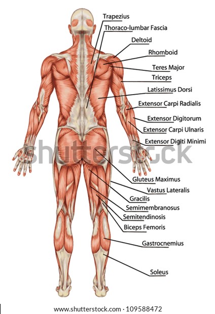
Anatomy Male Muscular System Posterior View Stock Vector Royalty Free 109588472 from image.shutterstock.com These muscles are also known as erector spinae (spinal erectors) or erector trunci (truncal erectors) since they specifically describe the primary function: Thoracic and abdominal muscles see online here the complexity of the musculoskeletal system is often a major issue for medical students. Human muscle system, the muscles of the human body that work the skeletal system, that are under voluntary control, and that are concerned with movement, posture, and balance.broadly considered, human muscle—like the muscles of all vertebrates—is often divided into striated muscle (or skeletal muscle), smooth muscle, and cardiac muscle.smooth muscle is under involuntary control and is. Head, neck and anterior torso muscles. It is situated on the right and left the. Superficial muscles of the torso male and female anatomy 1. It inserts onto the inferior border of the 12th rib. Orientation and landmarks to memorize.
Torso posterior muscles model, trapezius, deltoid, triceps brachii, latissimus dorsi, gluteus maximus, biceps femoris, deltoid, infraspinatus, semitendinosis are some names of body part. Orientation and landmarks to memorize. Figurative anatomy muscles of the torso. How do muscles move bones? Learn about torso muscles with free interactive flashcards. This video is for educational purposes only!!! The intermediate layer contains the internal oblique, and the deep layer contains the transversus abdominis. There are around 650 skeletal muscles within the typical human body. Muscle anatomy of the human body. Muscles of the lumbar spine of the trunk : Chest muscles function in respiration while abdominal muscles function in torso movement and in maintenance of balance and posture muscles of torso. As these muscles contract and relax, they move skeletal bones to create movement of the body. Skeletal muscle, posterior, unlabeled figure 8.16 250.
Captions with name and actions of the muscles have muscles of the torso. The intermediate layer contains the internal oblique, and the deep layer contains the transversus abdominis.

